
Hip Labrum (Cartilage) Injuries
Anatomy & Function:
The labrum is a rim of cartilage that surrounds the hip joint. It attaches to the socket of the acetabulum of the pelvis. The meniscus of the knee and the labrum of the hip are made up of the exact same type of cartilage.
The labrum increases the relative depth of the socket, which provides additional stability to the hip. This can be especially important in people who put their hips through extreme ranges of motion.
The labrum also acts a seal around the femoral head, to maintain fluid pressure of the hip joint.
Labrum tears:
The severity of symptoms from these injuries can vary. The “hip pain” 90% of the time is perceived as deep in the groin. Sometimes, the pain can radiate to the side or the back of the hip as well. Many times, those with a labral tear have pain and/or a feeling of catching in their hip, especially when going from sitting to standing. They may notice that they have to compensate to get in and out of cars. They may also have pain with squatting and exercise, especially with sports that involve cutting and changing direction. Many times, patients also complain of pain during sex.
Diagnosing a Labral tear:
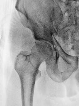
We can suspect a labral tear based on a person’s symptoms and history. Clinically, we can do provocative tests, such as the anterior and posterior impingement test, the McCarthy test, and the grind test, to further investigate. Many times, X-rays are very useful. Femoro-Acetabular Impingement (FAI), where the head of the femur is “out of round” can usually be seen on plain X-rays. With severe FAI, a labral tear can be inferred, due to the abnormal forces placed on the labrum by the unusually shaped “ball” of the femoral head.
MRI directly shows the cartilage of the hip. While a labral tear can often be diagnosed with a plain MRI, an MRI arthrogram, involving an injection directly into the joint, makes the diagnosis clearer if dye can easily be seen leaking from a labral tear.
Treatment/Management options for hip labrum tears:
The goal during conservative physiotherapy management of an acetabular labral tear is to optimize the alignment of the hip joint and the precision of joint motion. This can be done by-
Through gait (walking) and foot motion analysis, any abnormalities such as knee hyperextension causing hip hyperextension, walking with an externally rotated hip, or stiffness in the subtalar (in the foot) joint can be analysed and can be corrected through taping, orthotics or strengthening. Gait analysis may also uncover decreased hip abduction during both the stance and swing phase, as well as decreased hip extension during swing phase- characteristics that may be part of a hip joint stabilization strategy used by these people to compensate for deficient hip musculature functionality.
Additionally, education regarding modification of functional activities to avoid any positions that cause pain, such as sitting with knees lower than hips or with legs crossed, getting up from a chair by rotating the pelvis on a loaded femur, or hyperextending the hip while walking on a treadmill needs to be undertaken.
After addressing abnormal movement patterns, focused muscle strengthening work and recovery of normal range of motion, progression to more advanced training and functional exercises, sport specific if applicable, can be made.
Surgery:
The most common surgical procedure for a torn acetabular labrum is an excision or debridement of the torn tissue by joint arthroscopy. However, studies have demonstrated mixed post-surgical results. Fargo et al found a significant correlation between outcomes and presence of arthritis on radiography. Only 21% of patients with detectable arthritis had good results from labral surgery, compared with 75% of patients without arthritis. For a simple tear the surgery involves a bio absorbable suture anchor being placed over the tear to stabilize the fibrocartilaginous tissue back onto the rim of the acetabulum when the labrum has detached from the bone.
If surgery is performed, usually the first 6 weeks post-surgery are non or touch weightbearing only. Active and active assisted exercises are appropriate in gravity-minimized positions to maintain motion of the hip. Stationary bike, not recumbent bicycle, is appropriate; end range hip flexion should be done passively rather than actively. Rehabilitation protocols are currently based on surgeon and physiotherapists’ experience and can follow either labral debridement or repair guidelines, depending on the procedure performed.
I hope this detailed summary will help you or someone you know. As always, we are here at The Physio Nook to help out with any musculoskeletal disorders you may have, ankle or otherwise! Feel free to call us, email, or drop in for a great service.
Paul Woodward
Principal Physiotherapist
The Physio Nook.
The severity of symptoms from these injuries can vary. The “hip pain” 90% of the time is perceived as deep in the groin. Sometimes, the pain can radiate to the side or the back of the hip as well. Many times, those with a labral tear have pain and/or a feeling of catching in their hip, especially when going from sitting to standing. They may notice that they have to compensate to get in and out of cars. They may also have pain with squatting and exercise, especially with sports that involve cutting and changing direction. Many times, patients also complain of pain during sex.
Diagnosing a Labral tear:
We can suspect a labral tear based on a person’s symptoms and history. Clinically, we can do provocative tests, such as the anterior and posterior impingement test, the McCarthy test, and the grind test, to further investigate. Many times, X-rays are very useful. Femoro-Acetabular Impingement (FAI), where the head of the femur is “out of round” can usually be seen on plain X-rays. With severe FAI, a labral tear can be inferred, due to the abnormal forces placed on the labrum by the unusually shaped “ball” of the femoral head.
MRI directly shows the cartilage of the hip. While a labral tear can often be diagnosed with a plain MRI, an MRI arthrogram, involving an injection directly into the joint, makes the diagnosis clearer if dye can easily be seen leaking from a labral tear.
Treatment/Management options for hip labrum tears:
The goal during conservative physiotherapy management of an acetabular labral tear is to optimize the alignment of the hip joint and the precision of joint motion. This can be done by-
- Reducing anteriorly directed forces on the hip;
- Addressing abnormal patterns of recruitment of muscles that control the hip; &
- Instructing patients to avoid pivoting motions, especially under load, since the acetabulum rotates on a loaded femur, thus increasing force across the labrum.
Through gait (walking) and foot motion analysis, any abnormalities such as knee hyperextension causing hip hyperextension, walking with an externally rotated hip, or stiffness in the subtalar (in the foot) joint can be analysed and can be corrected through taping, orthotics or strengthening. Gait analysis may also uncover decreased hip abduction during both the stance and swing phase, as well as decreased hip extension during swing phase- characteristics that may be part of a hip joint stabilization strategy used by these people to compensate for deficient hip musculature functionality.
Additionally, education regarding modification of functional activities to avoid any positions that cause pain, such as sitting with knees lower than hips or with legs crossed, getting up from a chair by rotating the pelvis on a loaded femur, or hyperextending the hip while walking on a treadmill needs to be undertaken.
After addressing abnormal movement patterns, focused muscle strengthening work and recovery of normal range of motion, progression to more advanced training and functional exercises, sport specific if applicable, can be made.
Surgery:
The most common surgical procedure for a torn acetabular labrum is an excision or debridement of the torn tissue by joint arthroscopy. However, studies have demonstrated mixed post-surgical results. Fargo et al found a significant correlation between outcomes and presence of arthritis on radiography. Only 21% of patients with detectable arthritis had good results from labral surgery, compared with 75% of patients without arthritis. For a simple tear the surgery involves a bio absorbable suture anchor being placed over the tear to stabilize the fibrocartilaginous tissue back onto the rim of the acetabulum when the labrum has detached from the bone.
If surgery is performed, usually the first 6 weeks post-surgery are non or touch weightbearing only. Active and active assisted exercises are appropriate in gravity-minimized positions to maintain motion of the hip. Stationary bike, not recumbent bicycle, is appropriate; end range hip flexion should be done passively rather than actively. Rehabilitation protocols are currently based on surgeon and physiotherapists’ experience and can follow either labral debridement or repair guidelines, depending on the procedure performed.
I hope this detailed summary will help you or someone you know. As always, we are here at The Physio Nook to help out with any musculoskeletal disorders you may have, ankle or otherwise! Feel free to call us, email, or drop in for a great service.
Paul Woodward
Principal Physiotherapist
The Physio Nook.