
|
Most tissues in the body have healed completely in six to 12 weeks following an injury. However, many people have pain that lasts much longer than this. We know that the intensity of the pain you feel is not always associated with a corresponding amount of actual tissue damage. In some cases, there can be a severe amount of pain with almost no detectable damage. With this in mind, we explore some reasons why your pain might not be getting better, long after the tissues have healed.  You’re afraid of the pain?
Pain can mean many different things, for some of us pain can affect our ability to work or can be a symptom of a serious disease. What you believe about your pain can either amplify or reduce the symptoms you experience. If you feel that every time you experience pain you are causing more damage, you will naturally pay more attention to this and your nervous system will amplify the signals in an attempt to keep you safe. But if you understand the cause of your pain and know that while there is discomfort, you are not in danger of causing more damage, often the pain will feel less severe. This is one of the benefits of seeing a physiotherapist after your injury as they can help you to understand your pain, giving you more control over your recovery. You started moving differently after the injury? Immediately after an injury, it’s natural to change the way you move to avoid painful movements. After a while, these changed movement patterns can become a problem and actually begin to cause pain and discomfort on their own due to the altered stress patterns placed on your body. Correcting these adaptive movement patterns can often go a long way in reducing pain after an injury. You might not have noticed these changes and might need a physiotherapist to identify and help you to return to your usual movement pattern. You have lost muscle strength since the injury? While a certain amount of rest following an injury is helpful, if we stop moving altogether our muscles will lose strength. This can mean that our posture changes, we fatigue easier during our normal activities and we are more susceptible to further injury. Less movement also means we actually focus on the pain more when it does happen. Physiotherapists are able to advise you on the right types and amounts of exercise for you in the period following your injury. The pain has affected your lifestyle? When pain affects your ability to sleep, work and even concentrate, it’s not surprising that this can have a negative affect on your overall well-being and mental health. This can create a negative cycle of anxiety and depression that perpetuates and increases the experience of pain. If your pain is really getting you down, speaking to a mental health professional can actually be a valuable part of your physical recovery. Many other factors can obviously contribute to your neck pain not getting better, but these are four examples we commonly see. Contact your health professional for help with any persistent pain you may be experiencing.
0 Comments
What is it? Our knees are complex hinge joints, designed to provide stability from side to side and smooth movement forwards and back as you walk, kick and run. The patella, or kneecap, is a small bone embedded in the tendon of the quadriceps muscle that protects the knee and also provides extra leverage to the quadriceps, amplifying their strength. The patella moves up and down in a groove at the front of the knee as the knee bends and straightens. Usually this movement is smooth, with little friction, however, if something causes the patella to move in a dysfunctional way, the soft tissue between the kneecap and the knee can become irritated, causing pain in a typical pattern. This condition is often referred to as ‘runner’s knee’, PFJ syndrome or patellofemoral pain syndrome (PFPS).  What causes it?
The patella usually sits in a balanced position in the shallow groove at the front of the knee and moves easily without friction. The patella is attached to the quadriceps muscle at the top and connected to the lower leg via the patella tendon at the bottom. When the quadriceps contracts, this pulls on the patella and acts to straighten the knee. If one side of the quadriceps is stronger or tighter than the other, it can cause the kneecap to pull to one side and over time become irritated. There can be many factors that cause a muscle imbalance or weakness on one side of the quadriceps. In most people, the outer aspect of the quadriceps tends to be stronger and tighter than the inner muscle. Certain postures and leg positions require the outer muscles to work harder and the inside muscles to become less active. Lack of arch support in your feet or simply a physical abnormality of the knee can also place stress on the movement of the patella. What are the symptoms? This condition is characterized by pain felt on the inside or behind the patella with activities that require repetitive bending of the knee. There may be a sensation of crunching, clicking or grinding and some people report that their knee suddenly gives way. The pain is commonly felt when running, going up and down stairs or when doing squats and is generally relieved with rest. The pain may start as a small niggle and gradually become worse over time. How can physiotherapy help? The first step in effective treatment is to exclude any other conditions and have a physiotherapist confirm the diagnosis. Your physiotherapist is able to determine which factors are contributing to this condition, which could include poor posture, a lack of arch support in your feet or poor running technique. Once these factors have been identified, you will be provided with a specific treatment program to best approach your condition. PFJ syndrome usually responds well to biomechanical analysis and correction of any muscular weakness and imbalance. Having the correct shoes and orthotics can also make a huge difference. There are some short-term treatments, such as patella taping, dry needling, trigger point therapy and ultrasound, which may help alleviate symptoms quickly and keep you active while you address the other factors contributing to your pain. Stretching has long played an important role in the world of sport and fitness, with many athletes stretching religiously before and after exercise in hopes of preventing injuries. More recently, this practice has been called into question with many people wondering if stretching really makes a difference to athletic performance. The answer, like most things, is not black and white, as we explore a little in this article.  A brief introduction to stretching
Stretching is a type of movement that increases flexibility by lengthening muscle fibres to the end of their range. Stretching before and after exercise has been thought to reduce the risk of injury, improve athletic performance and reduce muscle soreness after exercise. The two most common types of stretching are static and dynamic stretching. Static stretching is when you lengthen your muscle and then hold that position for a period of time. Dynamic stretching uses movement and momentum of the body to stretch muscles to their end range, without holding the stretch at the end. What does the research say? Some research has suggested that static stretching before an activity can actually reduce power, strength and performance. However, these reductions were shown to be minimal and not noticed at all if the stretches were held for less than 45 seconds. It has also been found that stretching does improve flexibility but only for a short period of time. A few minutes after stretching, your joints move further, and with less resistance, so you may have improved flexibility immediately after stretching. Why stretch at all? One thing that is undeniable is that stretching feels great, with many people feeling more relaxed and reporting a rush of endorphins after a good stretching session. It is also difficult to test the long-term effects of stretching specific muscles showing abnormal tightness. A long-term static stretching routine will improve your overall flexibility, and this is thought to help prevent injuries, although the evidence is inconclusive. If you’re an athlete, the decision to stretch or not can be a personal one. A warm-up prior to intense exercise that includes some form of dynamic stretching is generally recommended for reducing injury risk, but of course is no guarantee. Strength and balance training may have a far greater impact on reducing injuries in the long term. Your physiotherapist is able to guide you on the best stretching advice for your individual activity and they may be able to identify some areas where improving your flexibility will help to reduce injuries and improve performance. Low back pain is one of the most common conditions treated by physiotherapists and if you are unlucky enough to have been a sufferer, you know that severe back pain can take over your life. With improved understanding, health professionals have come to identify some common myths about back pain that are inaccurate, misleading or even counterproductive...  Myth #1 – Discs can ‘slip’ out of place
Sitting between the vertebrae of the spine are soft discs that provide flexibility and shock absorption to the spine. In the past, many health professionals have told patients that these discs had ‘slipped’ as a way of explaining their pain to them. This is not entirely accurate, as these discs are actually very secure and rarely, if ever 'slip' out of place. Discs may bulge slightly or in some cases tear, however more often than not these injuries will heal without any permanent damage and exist in many people without causing any pain at all. Incorrectly thinking that a part of your spine has permanently ‘slipped’ out of place can cause you to move differently, which can create more pain and dysfunction in itself. Myth #2 – If you have low back pain, you should stay in bed When back pain strikes, our natural instinct is to rest, avoid movement and wait for the pain to pass. However, studies have shown that being active and performing targeted and gentle exercises can help improve low back pain. In fact, our impulse to stop moving and protect our spines can actually cause abnormal movement patterns and stress, leading to ongoing pain after the original injury has healed. If you are unsure of what kind of exercises you should be doing, your physiotherapist can help guide you with a targeted exercise program. Myth #3 – Severe pain means severe damage Pain that is severe, strikes suddenly and without warning can be a very scary experience. If this happens to you, you could be forgiven for assuming you must have sustained a very serious injury. The fact is, however, that the spine, being surrounded by nerves, is a particularly sensitive area of the body and pain in this area can be very strong without significant damage. A small ligament sprain or a muscle spasm can actually cause a large amount of pain, but it is common for intense symptoms to settle down quickly, even disappearing within a few days. In many cases, symptoms that last for longer than 2-3 weeks are caused by changes to your movement patterns in response to this pain and not the original injury itself. If you are suffering from back pain, the best person to see is your physiotherapist. They can help you to recover without any complications or side effects and help you safely return to your usual activities while also ruling out any serious damage that might need further investigation. Being active is one the most important aspects of a healthy lifestyle and there are many different ways to get your heart rate up. No matter what your choice of activity is, there is always some risk of injury. In this article, we have listed some tips to help you prevent accidents and injuries.  1. Choose the right footwear
The correct footwear can go a long way in protecting your feet and ankles from injury and can even prevent serious accidents such as falls. Every activity places different demands on your body and tailoring your footwear to suit these stressors is a great strategy for preventing injuries. For example, basketball players often wear shoes with support that extends above the ankles to help protect against ankle sprains, while hikers require thick and supportive soles to cushion and protect their feet. Wearing shoes that are too large or have poor grip can lead to slips and falls, particularly when exercising in the outdoors. Your physiotherapist can guide you with the correct choice of footwear for your chosen activity. 2. Pace yourself When you start to see improvements in your fitness and strength, it can be tempting to push your limits to see just how far you can go. The danger in this is that often your tissues are still adapting to the increased demands of your new exercise regime. Increasing your weights, training time or running distances by too much too soon can lead to major setbacks. Give your body time to adjust and progress in a slow and steady manner. 3. Check your form and posture Checking your posture in the middle of a workout is probably the last thing on your mind, however poor form is a leading cause of injury in athletes. As an example, lifting weights when your spine is not in its optimal position causes many low back injuries. Taking a second to check your posture before starting a lift is highly recommended. 4. Seek professional advice Coaches and trainers are able to help you spot vulnerabilities and share their knowledge, helping you get the most out of your chosen activity. Often it is easier to prevent bad habits from forming than it is to break them once they are already in place. Invest in the advice of an expert, they can help you to avoid injuries as well as reach your peak performance. Your physiotherapist is able to identify weakness in your training technique, biomechanical vulnerabilities, tight and/or weak muscles and can help guide you through your recovery if an injury does occur. However, prevention of injuries is always preferable to treatment whenever possible. There is no doubt that the human body can be very resilient. Short of regenerating new limbs, our bodies are capable of recovering from large amounts of damage, including broken bones. With this in mind, many people are happy to let nature take it’s course following an injury, thinking that seeing a physiotherapist will only act to speed up already healing tissues. The speed of recovery, however, is only one measure of healing and despite our body’s incredible capacity for repair, injury repair can be less than straightforward. Here are a few things about injury healing you may not have been aware of...  1. Scar Tissue is more likely to form without treatment.
Scar tissue can cause ongoing pain and stiffness in skin, muscles and ligaments. Physiotherapy can prevent excessive scarring from forming through advice regarding movement, massage and other hands-on treatment. 2. Your ability to sense the position of your body, known as proprioception, is often damaged after an injury and can be retrained. Impaired proprioception is a major factor in re-injury. If you’ve ever heard someone say “my knee/ankle/shoulder still doesn’t feel 100%” then this could be why. The good news is that with a specific exercise program, proprioception can be improved and restored. 3. Once healing has finished, your body may not be exactly the same as before. Following an injury, ligaments may be lax, joints may be stiffer and muscles are almost always weaker. While the pain may be gone, there might still be factors that need to be addressed to prevent more complicated issues in the future. 4. You may have picked up some bad habits while waiting for the injury to heal. While in pain, we often change the way we do things, this can lead to the development of poor movement patterns and muscle imbalances. Even though the pain has gone, these new patterns can remain and create further problems down the road. 5. Injuries don’t always heal completely. On occasion, injuries may not be able to heal completely on their own. The most serious example of this is a fracture that cannot heal if the bone is not kept still enough. Other factors that may prevent an injury from healing include poor circulation, diabetes, insufficient care of the injury and poor nutrition. Your physiotherapist can assess your injury and develop a treatment plan that will both restore you to the best possible function and prevent further injuries. Almost everyone will experience lower back and/or neck pain at some point in their lives, even if just in the form of a slight neck twinge after sleeping in an odd position. Spinal pain of the thoracic region (the upper & middle part of your back) is less common, however you might be surprised to discover how important this part of the body can be when it comes to pain and injury. 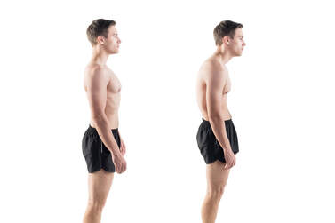 What is it?
The thoracic region refers to the part of the spine that is surrounded by the rib cage. It consists of 12 vertebrae with discs that sit between each of them. The thoracic spine isn’t an area that you might associate much with movement, however, this area can account for a surprising amount of flexibility, particularly in rotation. With joint attachments both between each side of the 12 vertebrae and a rib on either side, the thoracic spine has almost more individual joints than you can count. If each of these joints is not regularly moved through their full range they can tighten up and lose flexibility. This stiffness can become quite significant over time. Why is it important? Many people may not even notice this lack of movement, primarily because the neck and lower back provide much more range and can usually compensate for any loss of thoracic flexibility to complete everyday tasks. When there is no movement occurring in the thoracic region, this means that the structures of the joints in other regions are pushed closer to their limits of range, particularly during rotation. This results in more compression and stress on these joints and the structures surrounding them, such as nerves, blood vessels and muscles. Thoracic stiffness can be a significant risk factor for neck and lower back pain. This can also reduce the mobility of the chest wall, which can result in less efficient breathing mechanics and, in extreme cases, even reduced exercise tolerance. Thoracic spine movement is aslo important for normal shoulder function, as well as maintaining good posture. How can physiotherapy help? Your physiotherapist is able to assess your thoracic mobility and help you with treatments to improve your range, both with manual therapy and home exercises. They may even help improve your thoracic flexibility as part of a treatment plan for neck and lower back pain. There’s no doubt that doing the right thing for your health can be difficult. Whether that involves eating healthier, exercising more, flossing your teeth or even doing your physio exercises, the best choice is not always the most comfortable.  Once a habit has formed and become part of your routine, making the right decision for yourself can become a lot more straightforward. Getting to that point is easier said than done. Here are a few tips to help you reach your health goals...
1. Choose a friend to join you for accountability. One way to guarantee that you show up for an early morning gym session is to have a friend who you partner with. You might be comfortable sleeping in for yourself, but not so much when you have someone else you’re letting down. Sharing difficult tasks with a friend can make them more enjoyable and you can also set up competitions with them if you need a little extra motivation. 2. Combine tasks with an activity that you do like. Love a true crime podcast? The gym can be the time to catch the latest episode or even stream your favourite show. Anything you do that helps to increase the enjoyment of the activity you’re avoiding is going to help you get started. 3. Batch tasks to be more efficient. Are you trying to drink more water, floss and also complete your physio exercises? Why not do all three in the same sitting? Combining tasks can be a surprisingly effective use of time and a way to remind yourself to complete them. 3. Use positive reinforcement for good behaviour, rather than punishments. Many people will feel bad when they fail to reach their goals and try to restrict themselves in another area to compensate. It can be a much more effective motivation strategy to reward yourself after completing a task, rather than punish yourself for not doing it. 4. Get your foot in the door. Putting your shoes on can be the hardest part of going for a run. If you’re struggling to complete a task, sometimes all you have to do is tell yourself you only need to do five minutes. Once you’ve started quite often you’ll want to do more, but setting small goals can be the trick to getting started. Once a habit is in place, the easy part is to increase the intensity or length of time you’re doing it. It's believed that it takes 21 days of completing a task for a habit to be formed, where you will then do it automatically as part of your routine. For more tips and tricks on how to reach your health goals, speak to your physiotherapist. Chronic ankle instability is an instability affecting the ankle joint and its surrounding structures. It usually develops after a severe ankle sprain. However some people are born with less stable ankles, who may be generally extra flexible throughout their entire bodies. Approximately 20% of ankle sprains lead to chronic ankle instability due to the resulting changes in ligament support, strength, postural control, muscle reaction time and sensation.  What are the symptoms?
As well as being more susceptible to ankle sprains, people with chronic ankle instability may notice they are extra cautious during high-intensity activities, if walking or running on uneven surfaces or when changing directions quickly. They may experience a sense of weakness or "giving way" when weight-bearing. What are the causes? The primary causes of this condition are ligament laxity, decreased muscle strength of the muscles surrounding the ankle and reduced proprioception. Following an ankle sprain, ligaments can be stretched and slightly weaker. In more severe cases, they may have torn altogether, leaving the ankle structurally weaker. Without full rehabilitation, the surrounding muscles also become weaker, and studies have shown that balance and sensation of the ankle can also be reduced. This means that the ankle is more likely to be injured again, creating a vicious cycle leading to further instability. How can physiotherapy help? Physiotherapy treatment for chronic ankle instability focuses on improving strength, control and balance with a variety of techniques. This approach can significantly improve dynamic ankle stability and reduce the risk of future sprains. Physiotherapists can help patients to regain confidence and get back to their best performance. In some cases, taping or bracing for support can be used. However this can lead to dependence and further loss of strength and control if used unnecessarily. In cases of extreme ligament laxity or if physiotherapy fails, surgery to repair the damaged ligaments is considered. This is usually combined with a full post-operative physiotherapy rehabilitation program for greatest success. What are they? The calf muscles refer to a group of muscles at the back of the lower leg that act to point the foot and toes down, away from the body. They play an important role in walking and running. A tear or strain of these muscles occurs when some or all of the muscle fibres are torn or overstretched. This is a common injury that can affect anyone from elite athletes to recreational exercisers. 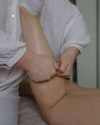 How do injuries happen?
Calf tears are often caused by sudden, forceful movements or overuse of the calf muscles, leading to the rupture or strain of muscle fibers. Common mechanisms of injury are a quick take off during sports or simply going for a long walk when not accustomed. Factors that may increase the risk of a calf tear are previous calf tears that have not been fully rehabilitated, tight or weak calf muscles, poor balance and poorly fitting footwear. What are the symptoms? Typical symptoms of a calf tear are sharp pain over the site of the tear, especially with movement, swelling, bruising, and difficulty walking or standing. The severity of the injury can range from mild muscle strain to a complete tear, which will determine the appropriate treatment approach. How can physiotherapy help? The first step in managing calf tears is accurate diagnosis by a medical professional, who is able to rule out other conditions that might mimic a calf tear. They can determine the extent of the damage and create a personalised treatment plan based on your specific needs. This ensures that the rehabilitation process addresses the root cause of the injury, leading to better outcomes. Reducing pain and inflammation is important in the first one to two days following the injury. The muscle may need support during this time, depending on the severity. Over time as the swelling and inflammation subsides, your physiotherapist will help to address any factors that contributed to the injury such as muscle weakness or imbalances. Calf tears often lead to stiffness and limited range of motion in the affected leg if not fully rehabilitated. Physiotherapists implement targeted stretching and range of motion exercises to restore flexibility and prevent the formation of scar tissue that may impede recovery. Gradually, the patient can regain the ability to move the calf muscle without pain or discomfort. Rehabilitation past this point will progressively challenge the calf muscles without causing further damage. Strengthening these muscles not only aids in the healing process but also reduces the risk of future calf tears. |
|
|
|