
|
The lower part of the leg, from the knee down to the ankle consists of two long bones that sit side by side- the thicker tibia and the thinner fibula. The bones are joined together by thick fibrous connective tissue called a "syndesmosis" and are firmly attached to each other with just a small amount of movement between them, allowing for a small amount of rotation of the ankle. A fracture of the fibula occurs when the bony tissue is disrupted or broken. It is a common injury and can occur at any part of the bone, depending on the mechanism of injury or the state of the bone. 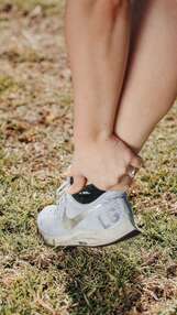 How does it happen?
A fracture of any bone can occur when the force applied to any point exceeds the strength of the bone at that point. However, as with all fractures, there are common patterns that are seen based on structural points of weakness in the bone and common patterns of movement. A few common ways that the fibula is broken are... Blunt force: If something hits the bone hard enough, it will break on impact. This could include being hit by a ball hard enough or being hit by a car, as this is the site where a car's bumper would reach. Impacts like this that have enough force will often break both the tibia and the fibula at the same time. Skiing accidents where skis hit something suddenly or get stuck can also cause the bones to break at the upper level of the ski boots. Ankle Sprain: When it comes to the fibula, the most common reason for the bone to be broken is during a severe ankle sprain. The ligaments that attach the outside of the foot to the fibula are so strong that when you twist your ankle badly enough, sometimes it is the bone that breaks. This is one of the most commonly missed injuries, partly because the fibula is not a weight-bearing bone. This means that after the initial pain and swelling have subsided, you can still walk on your foot without pain stopping you. It is important to have any severe ankle sprains imaged by X-ray to rule out any fibula fractures. What are the symptoms? In some cases, the symptoms of a fibula fracture will be unmistakable, with severe pain. Sometimes the skin will be broken and there will be bleeding. If the bone has been moved from its usual position, there will be a deformity such as a bump or dip under the skin. For smaller, undisplaced fractures, there will be pain over the bony aspects and a constant, deep pain that is worse when weight-bearing. What is the treatment? Physiotherapists are often the first to notice fractures caused by ankle sprains. Once a fracture has been confirmed, your medical team will decide on the best course of action to allow the bones to heal. This might include surgery to pin the bones together, casting or the use of a moon boot. Following a period of immobilisation, your physiotherapist can help you rehabilitate the surrounding tissues. This will include muscle strengthening, joint mobilisation, balance and control retraining, and a stretching program.
0 Comments
What is it? Shin splints are a painful condition of the lower leg, also known as Medial Tibial Stress Syndrome (MTSS), it is an overuse injury that causes pain along the inside of the tibia (shin bone). It occurs most commonly in runners, hikers and soldiers who march long distances.  What are the symptoms?
Shin splints are typified by persistent leg pain, usually the inside of the shin, halfway down the lower leg. The pain might be felt during exercise or directly after. Some people experience a dull ache over their shin that lasts for quite a while after exercise stops, while for others the pain may be sharp and fade quickly. The pain is often progressive, becoming worse with shorter distances. Eventually, shin splints can severely impact activity levels as the pain becomes too severe to continue exercising. Shin splints can be extremely painful and very disruptive to activity levels. As the pain usually starts gradually and progresses many people find themselves unable to continue training. Shin splints may progress to stress fractures if not diagnosed early and managed effectively. How does it happen? Shin splints are predominantly seen in runners who increase their distances quickly, often while training for an event. Activities that require repetitive weight-bearing of any kind, such as marching or high impact sports have also been shown to cause shin splints. Although the pathology of shin splints is unclear, studies have been able to identify certain risk factors that may predispose someone to shin splints. These include... · An quick increase in activity level; · Improper footwear and support; · Higher BMI; · Training on hard or uneven surfaces; · Tight calf muscles; · Flat feet; · Increased external rotation range of the hips; · Females are more likely to develop shin splints than males; · Prior history of shin splints. How can physiotherapy help? The first step for your physiotherapist will be to address any contributing factors to your injury and help to adapt your training program to a level that is optimum for you. A period of relative rest may be recommended along with a targeted strengthening and stretching program for any weak or tight muscles. Switching to low-impact activities such as swimming, cycling and yoga may also help to maintain fitness during recovery. Your running technique may be analyzed and any training errors corrected. When getting back into your training routine, it is usually recommended that distances are not increased by more than 10% per week as this allows the tissues of the body to react to the increased demands and adapt accordingly. Osteitis Pubis is a medical term used to describe sports-related groin pain. Osteitis means ‘bone inflammation’, while pubis refers to the specific bone that is affected: the pubic bone. Osteitis pubis is usually an overuse injury that can sometimes be triggered by a specific event. It is characterized by pain deep within the front of the pubic bone, caused by inflammation. The area of the pubic bone affected is specifically known as the pubic symphysis. This type of injury is common in load-bearing athletes such as runners. Other people commonly affected include soccer players and footballers, due to their frequent kicking actions. 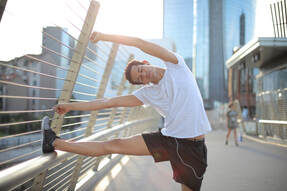 How does it happen?
Instability around the pelvic region is the primary cause of Osteitis Pubis, particularly if the instability occurs at the connection between the two sides of the pubic bones at the front of the body. The pelvis carries the weight of the upper body and is responsible for providing stability when walking, running and kicking. This means that the joint can become irritated and inflamed. What are the signs and symptoms? Osteitis pubis is aggravated by weight-bearing activities, with running and kicking being the two main culprits. Pain is usually experienced on one side, however both sides can be affected. The pain is usually located at the front of the pelvis and may progress into the hip and groin area as it becomes more severe. Sufferers of Osteitis Pubis may have a history of a previous groin strain, as well as lower back pain. They may also have a history of a sports hernia in the hip area. As with most inflammatory conditions, the pain may be worse when in use, better when resting, and worse overnight into the morning. How can Physio help? Your physiotherapist can help this condition in several ways and will aim to get you back to your pre-injury sporting level. During the assessment, your physio will look at many different things to determine the cause of the condition. Muscle length, muscle strength and muscle control will all be assessed. Your posture in standing, walking and running can also be assessed to determine any irregularities. Your physio will ask you to rest from sports for some time to allow some bony healing to occur. They will then progress you through a rehabilitation program aimed at getting you back to sport. This rehab program will retrain your muscles to stabilise the pelvis when walking, running and kicking. The muscles will also need to have relatively equal flexibility to help stabilize the pelvis. Your physio will give you specific exercises to target the strength and flexibility of these muscles. Finally, your physio will progress you to running or kicking, and allow you to gradually return to sport over a 3 to 6 month period. The benefits of keeping active may seem obvious, yet it can't hurt to be reminded of the many ways exercise can improve your life. Here are a few of our favourite reasons to get moving...  1. Exercise improves energy levels.
Improving your fitness means your body is capable of achieving more for the same energy expenditure. While doing exercise can make you tired in the short term, regular improvements to your fitness will help you get more out of your body each day, with seemingly less effort. 2. Exercise can help to reduce stress. If you are stuck in a state of stress or anxiety, exercise can help you move out of it into a calmer and more relaxed state, improving your mood, concentration and sleep. There are now many studies to support this. 3. Exercise and hobbies can help you build connections and community. Making new friends as an adult can be surprisingly difficult and the importance of connection and community is being recognised more and more as being essential for overall wellbeing. Being part of a team, group or club is a great way to build confidence and meet friends as well as keeping active. 4. Exercise keeps your muscles, tendons, joints and bones healthy. Our bodies are often compared to machinery or car parts. However, there are some crucial differences between our bodies and machines, including the fact that our bodies respond to exercise by becoming stronger and healthier, rather than being worn out. As an example, one of the best ways to prevent osteoporosis is through regular high impact activity, which stimulates bone growth. 5. Exercise can help to reduce injuries. Similar to the previous point, tissues that are used regularly are stronger, more elastic and are less likely to tear or break when under stress. Regular exercise is the best way to keep your body in a healthy state and prevent injuries. Finding the right exercise for you can be tricky, your physiotherapist can help you with suggestions based on your ability and skillset if you need some help. What is it? The scaphoid is a small bone in the wrist that connects the radius bone in the forearm to the hand, situated near the thumb. Scaphoid fractures are a relatively common wrist injury and are commonly misdiagnosed, as the pain can be similar to a sprained wrist, even when the bone has been broken. Scaphoid fractures are notorious for complications in healing due to low blood supply to the area and how easily their diagnosis can be missed. 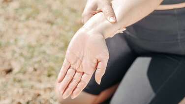 How does it happen?
A scaphoid fracture is often caused by a fall on an outstretched hand (FOOSH) or a direct blow to the wrist. It is more common in young adults than in children and the elderly. What are the symptoms? Symptoms of a broken scaphoid include wrist pain, swelling, bruising or discolouration of the skin over the injured area and difficulty moving the wrist or hand. As the swelling subsides you might notice pain at the base of the thumb when opening jars or gripping objects. There may also be a deep, dull ache in the wrist that doesn't settle easily. How is it diagnosed? If you suspect that you have a scaphoid fracture, you should consult your physiotherapist or GP who will refer you for an X-ray to confirm if the bone is broken. Occasionally scaphoid fractures will not show up on an X-ray, so if the findings are negative yet your medical team still suspect a fracture, they may wait a week then X-ray again or send you for an MRI or CT scan to double-check. Though these fractures can often be treated without surgery, doctors may recommend surgical intervention for more severe cases or if the bone is not healing well enough on its own. How can physiotherapy help? If you have a scaphoid fracture, your doctor will likely prescribe a splint or cast to ensure the wrist is kept still until healing is complete, usually for a minimum of six weeks. Healing times will vary depending on which part of the bone has been broken. Following the removal of the cast or splint, there is often residual pain, stiffness and muscle weakness. Your physiotherapist can help you restore any deficits as well as resolve any other aches or pains that may have resulted from altered biomechanics. What is it? A broken collarbone, or clavicle, is one of the most commonly broken bones in the body. The clavicle connects the front of the ribcage to the shoulder and is the only bony connection the arm has to the rest of the body. Many muscles attach to the clavicle, including the deltoid and pectoralis Major.  How does it happen?
The most common way for this injury to occur is through a fall onto the shoulder. This can happen from a simple fall or sports such as mountain biking or football. It is a common childhood injury but can happen at any age. What are the symptoms? Usually, a broken collarbone will cause moderate to severe pain over the broken area. The patient may have heard or felt a popping or cracking at the time of the injury and there may be an ongoing grinding or creaking with movements of the upper arm. If the skin is not broken there may be bruising and swelling over the painful area. What is the treatment? While very severe cases can be surgically fixed, more often a broken collarbone will be allowed to heal naturally with rest and monitoring. By supporting the arm in a sling and providing pain relief the arm will mend on its own. As with most fractures, there are often other injuries that need to be dealt with at the same time. There are many important structures near the collarbone that can be damaged, including muscles, nerves and blood vessels. In very severe cases, the lung tissue under the collarbone can be damaged causing the lung to collapse. Physiotherapy and recovery: Once a treatment plan has been decided by your medical team, your physiotherapist can help you to return to your pre-injury strength and mobility with a full rehabilitation program. What is a stress fracture? A stress fracture is a microscopic fracture of the bone that is often not picked up on X-ray. If left untreated, a stress fracture can cause significant disability and develop into a full fracture, possibly even requiring surgery. The majority of stress fractures occur in the lower limb, being particularly common in the hip, shins and foot at points where the most force passes through when weight bearing. Most stress fractures are overuse injuries and are common in long distance runners. 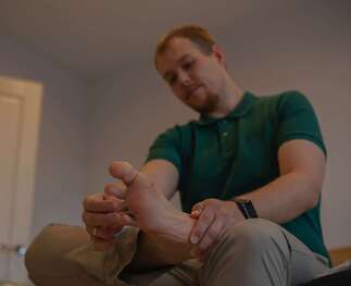 What are the symptoms?
As with many overuse injuries, the pain of a stress fracture starts gradually, beginning with pain during or after activity or sometimes the morning after. If activity continues without modification, the pain will gradually increase. Eventually most people are unable to maintain their usual activity level. Stress fractures are common in runners and military personnel who are required to march for long periods. A stress fracture will be more likely to occur in a person who has weaker bone strength, such as someone with osteoporosis, which is itself affected by many factors such as adequate calcium intake, vitamin D deficiency and a history of inactivity. How are stress fractures treated and how long will it take to get better? Stress fractures can easily be mistaken for other conditions such as shin splints. As the fracture is often too small to show up on X-ray, definitive diagnosis can be made using MRI, CT or bone scan. After diagnosis, the most important part of treatment will be resting the area to allow the bone to heal before resuming activity. Stress fractures usually need at least 6 weeks to recover fully. Some areas of the body have poor blood supply, which makes healing more complicated. For example, stress fractures of the navicular bone of the foot may need to be kept still and placed in a boot or cast for a period of time to heal properly. Other aspects of treatment will involve correcting any factors that contributed to the original injury. There is some evidence that unsupportive footwear is a risk factor, along with poor biomechanics and weak muscles that provide inadequate support to the skeletal system during activity. Speak to your physiotherapist if you suspect you may have a stress fracture or if you want to know more. Osteoporosis is a widespread condition characterized by low bone mass or density. It is primarily a metabolic disorder related to age and general health with a variety of risk factors and causes. The most common and well known consequence of osteoporosis is weakened bones that can break from small forces that would usually be harmless. In osteoporosis, both the matrix of the bone (similar to scaffolding) and the density of the bone are affected. While bone seems like a static part of our body, it is continuously laid down and removed by our bodies. In osteoporosis, there is an imbalance between the growth and reduction in the bone so it becomes progressively weaker. As such, it is a progressive disorder that worsens with age. While the disease process might begin much earlier, symptoms are usually only noticed over the age of 50. 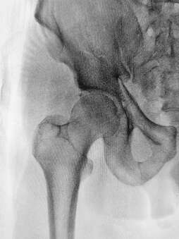 What are the signs and symptoms?
Often called a silent disease, many people with osteoporosis will have no idea that they have it, as there are no visible symptoms. Sometimes the first sign that an individual has osteoporosis is when the first bone breaks. Unfortunately, these bones are also slower to heal than healthy bones which can lead to ongoing complications. Broken bones are not the only symptom of osteoporosis, as bones lose density and strength they can also become compressed and develop wedge fractures under the weight of the body. When the spine is affected by osteoporosis, people may develop a hunched or stooped posture, which can itself lead to respiratory issues and place pressure on the internal organs. Osteoporosis can severely impact a person's mobility and independence, which can have a distressing impact on their overall quality of life. What causes it? As a metabolic disorder, osteoporosis can be caused by any process that interferes with the body's ability to maintain bone density. This includes gastrointestinal conditions that prevent adequate absorption of calcium (which is required for bone growth); lack of dietary calcium or low levels of vitamin D, which are essential for the absorption of calcium. Some medications can contribute to bone loss as an unfortunate side effect, especially if they are taken for a long time or in high doses. An example is the long-term use of steroids which can be prescribed to reduce inflammation. Inactivity can also predispose a person to osteoporosis as bones respond to force and weight bearing by building more bone. Having a sedentary lifestyle or choosing activities with low levels of impact can mean that without the weight bearing stimulus to make bone, bones are less dense over time. Osteoporosis can occur in elite cyclists and swimmers, who are more likely to develop the condition if they don't include weight-bearing activities such as jogging in their training program. How can physiotherapy help? Physiotherapy can help you to improve your overall bone health, and avoid or recover from fractures. Physiotherapy exercises can direct you to safely increase your weight-bearing, which can help build bone mass. Balance training is also an important factor as this can reduce your risk of falls. Your physiotherapist can also help you to adjust your lifestyle, at home or at work, to protect your bones and improve your posture, all of which are helpful in the overall management of osteoporosis. |
|
|
|