
|
Chronic ankle instability is an instability affecting the ankle joint and its surrounding structures. It usually develops after a severe ankle sprain. However some people are born with less stable ankles, who may be generally extra flexible throughout their entire bodies. Approximately 20% of ankle sprains lead to chronic ankle instability due to the resulting changes in ligament support, strength, postural control, muscle reaction time and sensation.  What are the symptoms?
As well as being more susceptible to ankle sprains, people with chronic ankle instability may notice they are extra cautious during high-intensity activities, if walking or running on uneven surfaces or when changing directions quickly. They may experience a sense of weakness or "giving way" when weight-bearing. What are the causes? The primary causes of this condition are ligament laxity, decreased muscle strength of the muscles surrounding the ankle and reduced proprioception. Following an ankle sprain, ligaments can be stretched and slightly weaker. In more severe cases, they may have torn altogether, leaving the ankle structurally weaker. Without full rehabilitation, the surrounding muscles also become weaker, and studies have shown that balance and sensation of the ankle can also be reduced. This means that the ankle is more likely to be injured again, creating a vicious cycle leading to further instability. How can physiotherapy help? Physiotherapy treatment for chronic ankle instability focuses on improving strength, control and balance with a variety of techniques. This approach can significantly improve dynamic ankle stability and reduce the risk of future sprains. Physiotherapists can help patients to regain confidence and get back to their best performance. In some cases, taping or bracing for support can be used. However this can lead to dependence and further loss of strength and control if used unnecessarily. In cases of extreme ligament laxity or if physiotherapy fails, surgery to repair the damaged ligaments is considered. This is usually combined with a full post-operative physiotherapy rehabilitation program for greatest success.
0 Comments
Ankle sprains are extremely common, however this doesn’t make them easy to cope with when they happen to you. If you’ve ever spent two weeks hobbling around on crutches after a bad twist, you’ll understand just how painful and difficult they can be.  What are they?
An ankle sprain refers to an overstretch or tear to the ligaments of the ankle. Commonly, a person will roll their ankle inwards and tear the ligament on the outside. Occasionally, the ankle will twist outwards and the ligaments on the inside of the ankle are torn and even less commonly, the fibres of the ligament that hold the two bones of the lower leg together can tear (this is a "high ankle sprain"). A sprained ankle will usually be painful, swollen, bruised, difficult to walk on and in some cases unstable. How does it happen? Ankle sprains can occur from something as simple as putting weight onto your leg when you think your foot is flat even though it’s not. The most typical pattern is of a person jumping and landing on the outside of their foot or simply slipping and twisting their ankle. A sprained or twisted ankle is one of the most common injuries presented to hospital emergency departments around the world. A medical or health professional should assess any ankle sprain. However, there are some guidelines to help decide if a sprained ankle needs an X-ray... 1. You are unable to put weight on the ankle immediately after the injury. 2. You are unable to take more than 4 steps immediately after the injury. 3. You have pain on the bony edges of the outer foot and ankle. How long do sprains take to heal? Depending on the severity of the tear, from one to six weeks. Your physiotherapist is able to help with recovery and ensure nothing slows down the healing process. Following any injury, joints may remain a little stiff and lose strength and control. Even though the injured tissues have healed, the ankle doesn’t move quite the way it used to. This means that your risk of twisting it again is higher than before the injury. How can physiotherapy help? Correct rehabilitation can help to speed up your recovery and prevent recurring injuries. As well as providing support to the unstable ankle, your physiotherapist will help you to strengthen any weak muscles and restore balance and control through exercise. They are also able to correct any abnormal movement of the joint following the injury. Strains and sprains are words that are used almost interchangeably when describing injuries, however they each have quite distinct meanings. The most straightforward explanation is that a “strain” refers to an injury in a muscle or tendon, while a “sprain” refers to an injury to a ligament. Here we describe what that means and how we treat strains and sprains differently.  Ligaments are fibrous tissues that connect and hold bones to other bones at your joints. These are very strong parts of your anatomy and provide large amounts of support and stability to the body.
Some ligaments are so strong that sometimes a bone will break before the ligament will tear. When ligament fibres do tear, the nearby joint can feel unstable as it has lost some of its structural support. A torn ligament will usually become painful and swollen, it may appear red and warm to touch and occasionally there will be some bruising. The pain will be worse with movement or if the ligament is placed under more stress. Occasionally, if a ligament has torn all the way through, the pain will not be as severe as it is with a partial tear. Your physiotherapist can grade the severity of a ligament sprain, which will help guide treatment and expected recovery times. Muscle strains are easy to confuse with ligament sprains, however, there are a few tell-tale differences. Following a muscle tear, it is more likely that you’ll feel weakness rather than instability. The pain will also be isolated over the muscle, rather than near a joint. An injury to a ligament will be tender over the site of the ligament and special tests can be done to test for any joint laxity. Treatment is also slightly different as sprains will need more support and will sometimes even need to be taped or braced, whereas muscle strains will benefit from gentle movements earlier. In both cases, following the basic principles of rest, ice, compression and elevation is great advice in the early stages of any injury. Applying heat is not recommended until at least two or three days after the injury. It is important to seek a professional opinion when recovering from both a strain and a sprain. It is very easy to re-injure an area while it's healing if undertaking too much activity too early and without correct rehabilitation. Speak to your physiotherapist for more information. If you've ever started a new hobby or activity and noticed your balance isn’t quite up to scratch, it can be quite a disturbing discovery. Balance is an important part of many activities and if it's not being challenged regularly, it's easy for it deteriorate without you noticing. 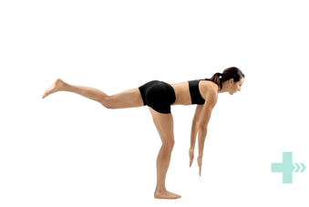 What is balance?
Keeping your balance refers to a state where your centre of gravity is maintained over your base of your support, preventing you from falling. Your body is always working hard to keep this equilibrium without you realising it. Balance is controlled by many systems that work together, including the visual, vestibular, proprioceptive and musculoskeletal systems. What is proprioception? Proprioception refers to the awareness of your body’s position in space. The central nervous system gains sensory input from the muscles, skin and tendons and interprets this information, creating a sense of where your body is positioned. This is how you know your foot is flat and ready to take your weight when you step, without needing to look at it. You may not have heard of proprioception before, but it is vital to keep you from falling and can be improved. How can I test my balance? Your physiotherapist is able to assess your balance more extensively, however here are a few quick tests you can do at home to see if your balance can be improved. Stand with two feet together and close your eyes. Try again, this time standing on one foot with your eyes open. Close your eyes only once you have found a steady posture with your eyes open. To increase difficulty, stand on an uneven surface, like a pillow on the floor. Aim to balance for at least 30 seconds in each of these postures. If you can't have a chat with your physiotherapist and see if your balance can be improved. They will also be able to offer you some practical tips on how to reduce falls and injuries. What is it? The AC (acromioclavicular) joint is a thick fibrous joint that connects the top of the shoulder blade to the outer end of the collarbone. The joint is required to be strong and supportive and is the primary way in which weight-bearing forces are transferred from the arm to the rest of the skeleton. The joint is connected by three strong ligaments, the acromioclavicular, corococlavicular and corocoacromial ligaments.  How does this injury occur?
The primary mechanism that will cause this joint and its ligaments to be injured is a force that separates the shoulder away from the collarbone, usually in a downwards direction. This can occur from a fall into the ground where the top of the shoulder hits the ground first, a rugby tackle or a fall onto an outstretched hand. As with all injuries, there are many variations in severity and a grading system has been developed to classify AC joint injuries. What are the symptoms? After an AC joint injury, there is usually immediate pain on the top of the shoulder, often with swelling and bruising. There is usually some loss of movement of the shoulder and pain from putting weight through the arm or carrying heavy objects. In severe cases, there is a visible lump on top of the shoulder known as a ‘step deformity’, where an obvious height difference can be seen between the top of the shoulder and the collar bone. There is frequently pain felt when reaching across the body, such as when putting on a seatbelt. To confirm the diagnosis, your physiotherapist can perform some clinical tests and sometimes an X-ray can help to grade the severity of the injury. The classification that would be given to you by your physiotherapist or doctor helps to determine the optimal course of action for each injury. There are different classification systems, some use four grades and the other six. Injuries with a smaller number of ligament fibres being torn are given a lower grade classification, going upwards as further damage is incurred. Injuries classified as higher grades will require surgical repair. How can physiotherapy help? The role of physiotherapy in this case is to ensure the joint is supported and given a chance to heal naturally while maintaining strength and movement of the shoulder girdle. This is done initially by providing support to the joint. You may need to have your arm supported in a sling or brace for some of this time, or your physiotherapist can show you some taping techniques to add support. Most AC joint sprains take around six weeks to fully heal, although some patients report ongoing shoulder problems years later. For this reason, a comprehensive rehabilitation program is very important. More severe sprains or total separations may require surgery to stabilise the joint and treat any associated fractures. Surgical repair will also require a proper rehabilitation program afterwards. The "socket" of the shoulder joint is surrounded by a ring of flexible connective tissue, known as a labrum. This labrum increases the stability of the shoulder while allowing the joint to stay flexible. The long head of the biceps tendon has an attachment directly into the labrum, which is often a point where injuries occur. A tear of the labrum can occur in many locations, however the most common is at the point where the biceps tendon attaches. Usually, this tear follows a typical pattern and is referred to as a superior labrum tear, anterior to posterior (SLAP tear).  How do they happen?
SLAP tears can be caused by trauma such as a fall onto an outstretched hand or can develop over time through repeated stress. Repetitive overhead activities such as throwing or painting can also gradually weaken the labrum over time and lead to a tear. What are the symptoms? Often if a SLAP tear develops over time, patients can be unaware they have sustained an injury at all and there is no significant impact on their pain or function. Pre-existing SLAP tears can however place more tension on the long head of the biceps tendon, leading to overuse disorders of that tendon as a secondary complication. When the tear occurs through a sudden action or trauma, symptoms can be more noticeable. Patients often notice pain deep in the shoulder joint with overhead shoulder movements, a feeling of weakness or a loss of power or accuracy with throwing activities. Some people may feel a popping or clicking sensation and occasionally the shoulder feels like it "gives way". In severe tears, the shoulder might feel unstable and be at increased risk of dislocation. How can physiotherapy help? Your physiotherapist can help diagnose a SLAP tear and send you for scans if needed. SLAP tears are graded by severity from 1 to 4 as a way to guide treatment. Physiotherapy is usually recommended as a trial for all tears before considering surgical repair and in many cases can effectively help patients return to their previous activities, symptom free. If physiotherapy is unsuccessful, surgical repair with a full rehabilitation program is then recommended. Surgery will either repair the tear or reattach the biceps tendon to the humerus (tenodesis). Following surgery, a period of rest in a sling is required before rehabilitation can begin. Essentially, the ankle consists of three bones, the tibia, fibula and talus, all held together by thick fibrous ligaments. The bottom parts of the tibia and fibula (shin bones) join together and surround the talus in such a way that it is able to rock forwards and back while providing stability and restricting the side-to-side movements. The ligaments holding the tibia and fibula together are large and thick, forming a syndesmosis. An injury to this area is known as a high ankle sprain. 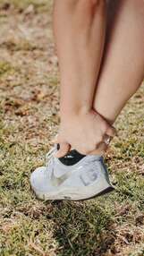 How do they happen?
A high ankle sprain can occur when your lower leg twists inwards while your foot is planted on the ground. The foot is typically pushed back and rotated outwards, putting excess pressure on the ligaments that hold the lower leg bones (tibia and fibula) together. This force can cause the syndesmosis to tear resulting in a gapping of the two bones, which can lead to significant instability of the ankle. This can happen simply from a fall, but more commonly while playing sports that involve running and jumping. This is also a common injury for downhill skiers. You are often unable to walk on your toes after this type of injury. What is the difference between a high and a low ankle sprain? A "normal" or low ankle sprain is a tear of the ligaments connecting the ankle to the foot, a syndesmosis tear (between the bottom of the tibia and fibula) is called a “high” ankle sprain. High ankle sprains are much less common than lower ankle sprains, accounting for only 1-11% of all ankle injuries. It can initially be difficult to tell the two injuries apart. To complicate things, a fracture of the ankle will also have similar symptoms. Your physiotherapist has a set of physical tests they can perform if they suspect a high ankle sprain. Ultimately imaging such as X-Rays or an MRI may be required to confirm the diagnosis. Why does correct diagnosis matter? High ankle sprains can take up to two times longer to heal than normal ankle sprains and require more immediate attention. Syndesmosis tears that are left untreated can result in chronic instability and pain, making them vulnerable to further injury in the future. Furthermore, if the separation of the tibia and fibula is severe enough, early surgery to fixate the injury with a screw or a "tightrope" to pull them back together may be required. What is the treatment? As mentioned, severe and unstable tears may require surgery, but most syndesmosis tears will need to be put into a supportive boot for around 4-6 weeks. Following this period a graduated rehabilitation program of strengthening, mobilization, balance, control and agility will need to be commenced before your ankle will be back to its pre-injury level of function. This can take up to 6 months in some cases. Anterior ankle impingement is a condition where repetitive forces compress and damage the tissues at the front of the ankle, causing pain and inflammation. It is a common injury that can affect people of all ages, however is usually seen in athletes of sports involving repetitive or forceful upward movements of the ankle, such as sprinting, landing from long jump, uphill and downhill running. 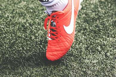 What are the symptoms?
Pain at the front of the ankle joint is the primary symptom of anterior ankle impingement. This can be felt as an intense, sharp pain occurring with ankle movements or a dull ache in front of the ankle following periods of exercise. Pain can also be felt when putting weight through the ankle while standing, walking or running. Night soreness, stiffness, swelling and reduced ankle flexibility are also common symptoms. How does it happen? Anterior ankle impingement is caused by traumatic or repetitive compression to the structures at the front of the ankle as the tibia and talus move towards each other during ankle movements. The tissues that are affected become damaged and inflamed, causing the pain typical of ankle impingement. Chronic inflammation can lead to further stiffness, exacerbating the impingement process. The most common risk factor for ankle impingement is a previous ankle sprain that was not adequately rehabilitated, as this can result in a stiff or unstable ankle. Another cause of impingement is the growth of small osteophytes or bony spurs around the ankle joint that press against the nearby soft tissues. These can be due to osteoarthritis or grow as a reaction to previous injury. Training errors, muscle tightness, unsupportive footwear and a hypermobile ankle have also been shown to be risk factors for anterior ankle impingement. How can physiotherapy help? Depending on the cause, mild cases of anterior ankle impingement usually recover in one to three weeks with rest and physiotherapy treatment. For more severe impingement, the ankle may require up to six weeks of rest and rehabilitation to recover. In rare cases, surgical intervention will be required to remove any physical causes of impingement such as osteophytes, to restore impingement free movement of the ankle. Your physiotherapist will first identify the cause of your ankle impingement and help you to choose the best course of action to reduce your symptoms. They are able to advise you on the appropriate amount of rest and provide stretches and exercises to restore strength and flexibility to the ankle. Mobilization techniques and range of motion exercises can also reduce stiffness of the ankle, restoring normal joint movement. Balance and proprioception exercises are also included to help prevent further ankle injury. Physiotherapy treatment is definitely the first step before considering surgery. If surgery is required however, your physiotherapist can help you to make a full recovery with a post surgical rehabilitation program. What is it? Your knee moves freely backwards and forwards; however, the thought of it moving from side to side probably makes you cringe. This is because the knee joint has sturdy ligaments either side of it that prevent sideways movement and we instinctively know that a lot of force would be required to shift it in this direction. The ligaments on either side of the knee are called the collateral ligaments and they each work to provide stability and restrict the knee’s movement into a sideways direction. The medial collateral ligament (MCL) is found on the inside of the knee and acts to prevent the knee bending inwards towards your other leg.  How does this injury occur?
The typical mechanism for this injury is a force that drives the lower leg inwards. This can occur from an awkward landing from a height, or when twisting with a foot fixed on the ground or from an external force hitting the outside of the knee, such as with a football or rugby tackle. What are the symptoms? MCL tears typically create pain and swelling quite specifically on the inside of the knee. The severity of the pain and swelling will be related to the amount of ligament fibres damaged. Larger tears may also make the knee feel unstable or loose. A grading system is used to classify the severity of the injury and help to guide treatment. Grade 1 indicates that a few ligament fibres have been torn and grade 3 is used for a complete tear of the ligament with associated joint laxity. Very severe MCL tears may also involve injury to the medial meniscus and anterior cruciate ligament (ACL) and can require surgical repair. However, most MCL sprains can be managed well with physiotherapy. Grade 1 and 2 MCL sprains take between 3-8 weeks to fully heal and a complete rehabilitation program is strongly recommended to prevent future injury. How can physiotherapy help? In the early stages of the injury, treatment is focused on pain and swelling management, while allowing the body to start the healing process. This is best managed thought the R.I.C.E. principles (Rest, Ice, Compression and Elevation). Following any injury, it is natural for muscles to waste a little and the damaged tissues to lose what we call proprioception, the ability to sense their position in space. This loss of muscle strength and proprioception can contribute to future injury if not restored with a proper rehabilitation program. Physiotherapy also aims to restore movement to the joint and support the ligament while healing to ensure that it is strong and healthy, and the scar tissue forms in an organized fashion, which makes the new ligament as strong as it can be and protects against future tears. What is the labrum of the shoulder? The shoulder is a remarkably mobile joint, however this flexibility comes with the cost of less stability. The glenohumeral joint, where the upper arm meets with the shoulder blade is a ball and socket type joint. The surface area of the ‘socket’ part of the joint (the glenoid fossa) is much smaller than the ball part of the joint (the head of the humerus). A fibro-cartilaginous ring called a labrum surrounds the edge of the glenoid fossa which acts to increase both the depth and width of the fossa. This labrum provides increased stability and is also the attachment for a part of the biceps muscle via a long tendon. The labrum can provide flexibility and stability that a larger glenoid fossa might not be able to, however being a soft structure it is prone to tearing which can be problematic. 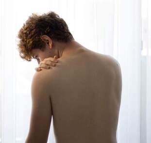 What causes the labrum to tear?
The most common way the labrum is torn is through a fall onto an outstretched arm or through repetitive overhead activities such as throwing or painting, as the repeated stress on the labrum can cause it to weaken and tear. Suspected labral tears can be diagnosed in a clinic by your physiotherapist through a series of tests, however, an MRI is usually required to fully confirm the presence of a labral tear. Labral tears are classified into different grades, which are determined by their location and severity. This grading is used as a guide to help determine the correct treatment. What are the symptoms of a labral tear? A labral tear is often associated with other injuries, such as a rotator cuff tear, which can make the clinical picture a little confusing. Commonly there will be pain in the shoulder that is difficult to pinpoint and the pain will be aggravated by overhead and behind the back activities. Severe labral tears can lead to instability and can also be related to dislocations of the shoulder. How Can Physiotherapy Help? The severity and grade of the labral tear will guide treatment. Smaller tears can be treated with physiotherapy that is aimed at increasing strength and control of the shoulder. Other tears may require surgical repair after which physiotherapy is an important part of treatment to rehabilitate the shoulder. |
|
|
|